No products in the cart.: $0.00
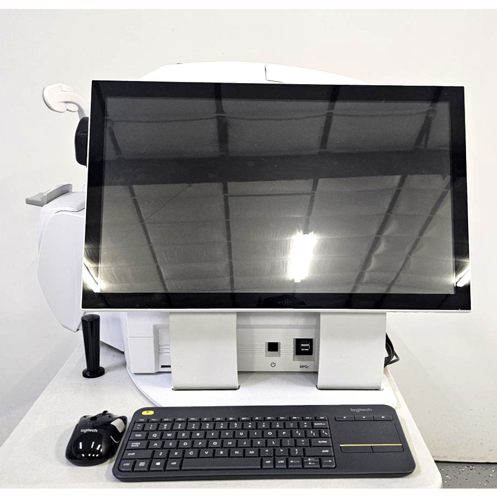
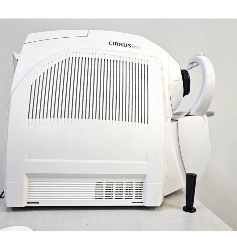
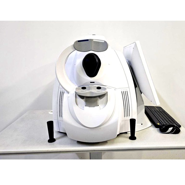

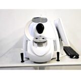
Carl Zeiss CIRRUS 6000 OCT Imaging System
The Carl Zeiss CIRRUS 6000 OCT Imaging System is a professional ophthalmic imaging solution that delivers high-resolution cross-sectional images of the retina, optic nerve, and anterior segment. Its fast scanning, advanced analysis software, and comprehensive reporting capabilities help eye care professionals evaluate ocular health, monitor disease progression, and support accurate clinical decision-making.
Description
Carl Zeiss CIRRUS 6000 OCT Imaging System – High-Resolution Optical Coherence Tomography
The Carl Zeiss CIRRUS 6000 OCT Imaging System is an advanced optical coherence tomography (OCT) device designed for comprehensive ophthalmic imaging. It produces detailed cross-sectional images of the retina, optic nerve, and anterior segment, helping eye care professionals evaluate ocular structures with exceptional clarity.
Designed for ophthalmology clinics, hospitals, and eye care centers, the system offers fast image acquisition, high-resolution scanning, and user-friendly software to support efficient diagnosis, treatment planning, and long-term patient monitoring.
Main Benefits
High-resolution retinal imaging
Fast image acquisition for efficient workflow
Supports comprehensive eye examinations
Detailed visualization of retinal layers
Assists with diagnosis and treatment planning
Reliable patient follow-up and comparison
Easy-to-use interface with automated analysis
Consistent imaging for clinical documentation
Key Features
High-definition optical coherence tomography
High-speed scanning technology
Detailed retinal and optic nerve imaging
Anterior segment imaging capability
Wide range of retinal scan protocols
Automated image analysis software
Thickness and structural measurements
Patient comparison and progression tracking
Integrated patient database
High-quality clinical reporting
User-friendly touchscreen interface
Digital image storage and export
Key Specifications
Product Type: Optical Coherence Tomography Imaging System
Imaging Technology: Spectral Domain Optical Coherence Tomography (SD-OCT)
Imaging Areas:
Retina
Macula
Optic nerve head
Retinal nerve fiber layer
Ganglion cell analysis
Anterior segment
Scan Resolution: High-resolution cross-sectional imaging
Scan Speed: High-speed image acquisition
Display: Integrated high-resolution monitor
Software: Clinical analysis and reporting
Patient Database: Integrated management system
Connectivity: Digital image export and network compatibility
Clinical Use: Ophthalmic diagnosis and patient monitoring
Applications
The Carl Zeiss CIRRUS 6000 OCT Imaging System is ideal for:
Ophthalmology clinics
Eye hospitals
Vision care centers
Retina specialty clinics
Glaucoma assessment
Macular disease evaluation
Diabetic eye examinations
Age-related macular degeneration monitoring
Routine eye health assessments
Clinical research institutions
What is the Carl Zeiss CIRRUS 6000 OCT Imaging System used for?
It is used to capture detailed images of the retina, optic nerve, and other eye structures to assist in diagnosing and monitoring various eye conditions.
What does OCT imaging provide?
OCT provides high-resolution cross-sectional images that help clinicians evaluate retinal layers and other ocular structures.
Who can benefit from this system?
Ophthalmologists, retina specialists, glaucoma specialists, optometrists, hospitals, and eye clinics can benefit from its advanced imaging capabilities.
Can the system monitor patient progress?
Yes. The software allows clinicians to compare previous and current scans to monitor changes over time.
Is the imaging procedure non-invasive?
Yes. OCT imaging is a non-invasive examination that captures detailed images without contacting the internal structures of the eye.